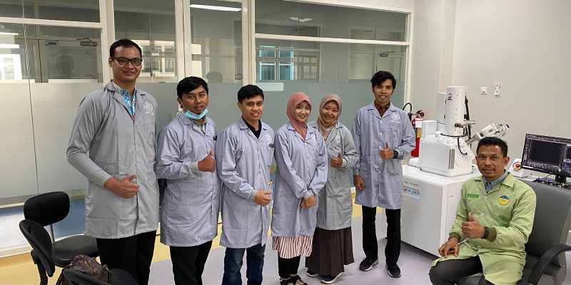
One of the widely-used research tools in current scientific research activities is the utilization of electron particles. The observation of electron particles through a microscope instrument provides valuable information and data regarding the ultrastructure of the observed samples, including surface topography, composition, and even elemental composition mapping. To keep up with the advancements, deepen their knowledge, and apply this instrument effectively, three members of the Plant Development Structure Laboratory staff, namely Utaminingsih, S.Si., M.Sc., Novita Yustinadiar, S.Si., M.Si., and Dr. Wiko Arif Wibowo, S.Si., participated in a training program organized by LPPT (Laboratory for Research and Integrated Testing) UGM. The FE-SEM (Field Emission-Scanning Electron Microscopy) training took place on August 31st to September 1st, 2023, at the FE-SEM Laboratory section of LPPT UGM.
The training activities were divided into theory sessions delivered by Prof. Dr. Eng. Yusril Yusuf, S.Si., M.Si., M.Eng. and Yusuf Umardani, S.T., M.Eng., as well as practical sessions supervised by Yusuf Umardani, S.T., M.Eng., and Virginia Fahriza, S.Si. The theoretical discussions covered fundamental concepts of FE-SEM, distinctions from SEM, SEM+EDX test result analysis and mapping, as well as techniques for capturing high-quality images.
This training event was held in a hybrid format with a total of 34 participants from various institutions across Indonesia.The material presented during the training provided a deeper understanding and insight into the use of SEM instruments in biological research, including its current developments that utilize probe current for electron particle emission in FE-SEM. Interactive discussions revolved around the preparation of biological samples required before observation. This is because biological samples typically contain a significant amount of liquids, whereas SEM observations ideally require samples to be in a dry state. Consequently, specific preparation and techniques are needed, depending on the type of biological sample being used. “This training has been highly beneficial in enhancing our analytical knowledge of anatomical structures, and we hope it will ultimately improve the quality of research and education in the SPT laboratory,” said Wiko in response to the training activities.
The use of electron microscopes in biological research has been widely adopted and has aided in the structural analysis of various fields of biology, not only in botany but also in zoology, microbiology, ecology, and environmental sciences, as well as in biomedical research, forensic biology, biodiversity studies, and more. Sample analysis services for observations using FE-SEM are currently available at LPPT UGM and SEM at FKG UGM.


